Pigment Microscopy: A tool for the identification of pigments in works of art by Ruth Siddall
Being a pigment analyst and my interest, specifically, is the analysis of artists’ pigments. By which I mean pigments that have been used by a historical or contemporary artist in creating a painting or installation. Whilst this may at first seem obvious, it is an important point to make. I am not simply a collector of pigments, their use must have a context for me to find them interesting. That does not mean to say that I don’t collect pigments, I certainly do and have over a thousand specimens. However, my collection is a comparative one, used to help identify materials I encounter in different contexts and from different periods and locations. The purpose of this knowledge is to inform art historical studies further about artists’ practices and the trade and technologies used in the manufacture and procurement of historical artists’ materials. It can answer the questions about how pieces of art were made when they were made and whether or not they have been restored (or indeed, counterfeited).
As physical and chemical entities, pigments can be derived from animals, vegetables and minerals, and be both natural and synthetic. They can be analysed by a range of techniques. The main tool I use is microscopy and specifically polarising light microscopy (PLM). To be useful as a pigment, a material must have one main property; it must retain its colour when ground to a very fine particle size, typically less than 40 millionths of a metre (40 µm) diameter. Microscopy, alone in all other analytical techniques, allows one to observe these tiny particles and to be able to see what colour they are (Fig. 1). This brings huge advantages to this technique. Mixtures of pigments, for example, blue and yellow combined to make green, are obvious. The relative size and shapes of particles are also readily observed, allowing the microscopist to identify whether these were ground, are precipitates or even contain fossil material. The identification of rock chalk (calcium carbonate) in painting grounds is made possible by the presence of tiny fossils called coccoliths, which are invisible to the naked eye, indeed small enough not be broken up by grinding the chalk (Fig. 2). Their presence can indicate the source of the chalk as well as quickly allowing differentiation from synthetic carbonates such as lead white and from calcium sulphate-based gessos.
The optical properties of pigment particles can be observed in-plane polarised light (white light) and under cross-polarised light. Combinations of observed properties in both light sources can allow firm identification of materials present. The technique of PLM has been routinely used for over 100 years in disciplines including geology (mineralogy) and pharmacy. Both organic and inorganic materials can be identified allowing PLM microscopists to characterise pigments, extenders and fillers used by artists. A limitation is experience; there is no quick way to learn this technique and machine learning has yet to replace the human eye. Another limitation is black opaque pigments, to provide a secure identification, a pigment particle must transmit at least some light!
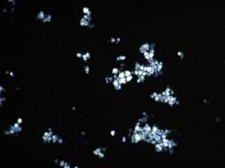
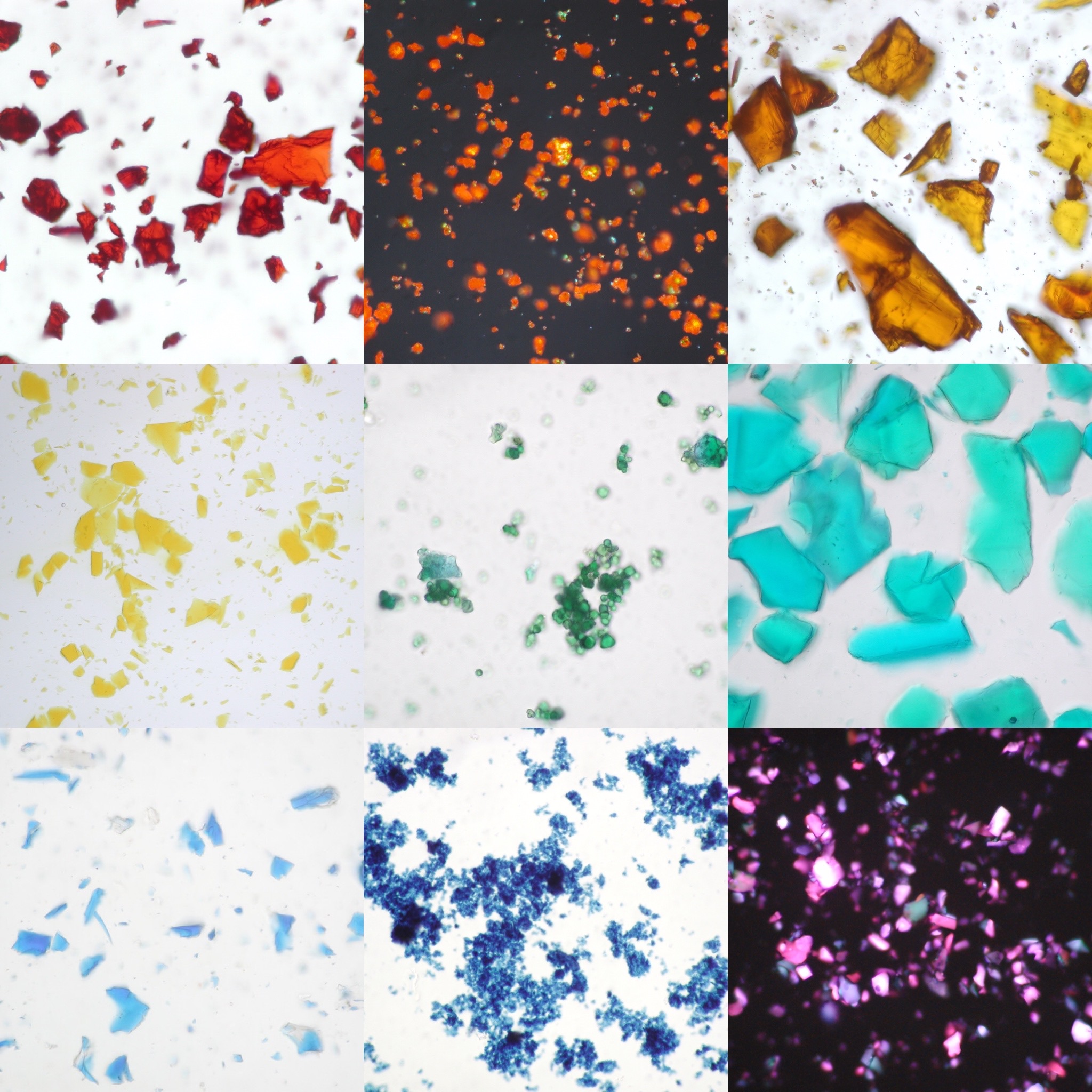
Figure 1. Pigments observed under plane polarised (bright field) and cross-polarised (dark field) light. Top row from left to right; vermillion, red lead, crocoite (that mineral form of lead chromate). Middle row from left to right; gamboge, cobalt zinc oxide green, verdigris. Bottom row left to right; lazurite (natural ultramarine), indigo, manganese violet. Angular particles such as those observed for gamboge and lazurite indicate a pigment that has been ground, whereas even-sized, small particles, such as those for red lead, cobalt zinc oxide and indigo indicate a chemical precipitate. Larger materials like the vermillion, crocoite and manganese violet have been allowed to crystallise and may be further ground down to produce pigment-size particles.
Figure 2. Natural chalk pigment, collected from geological outcrop. The pinwheel-shaped objects at centre-left are fossil coccoliths which are diagnostic of this material. This image is taken at 1000 x magnification using cross-polarised light.
Images © The Pigment Compendium 2004